
Structure-forming matrix
Consider formability, hydration and dissolution.
We develop dissolving microneedle formats for targeted skincare and new delivery applications. Our work brings formulation, array design, and manufacturing together around the needs of each product.
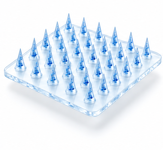
Dissolving microneedles are tiny, preformed structures made from water-soluble materials. They are arranged in arrays for controlled contact with the skin.
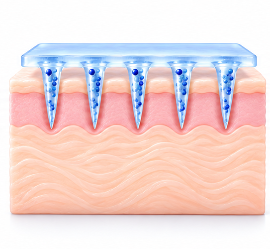
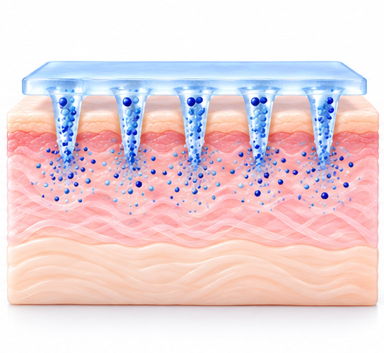
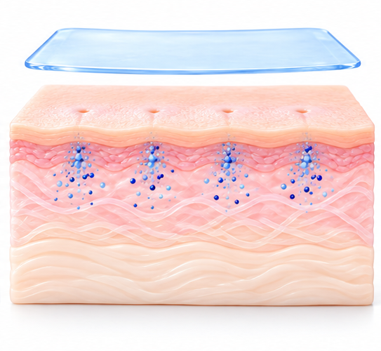
With the right material, geometry, and application method, the needles can pass through the outer skin barrier. Moisture then hydrates and dissolves the soluble material, releasing ingredients held within it.
Needle length, insertion depth, and ingredient distribution are separate measurements. Each product needs testing to determine where its ingredients go and how they are released.
 01
 02
 03
 04
Principle illustration, not to scale. It does not establish product-specific delivery depth or clinical outcomes.
Watch animation.
Microcrystal product animation · English version
Hyaluronic acid and sodium hyaluronate are key materials in our microcrystal products. The soluble matrix forms the needles and influences hydration, dissolution, and compatibility with other ingredients.
Formulation choices affect every step. Concentration and molecular properties influence flow and mold filling. Drying affects structural integrity, while contact with moisture affects dissolution.
A finished product also includes support and application components. Backings, adhesives, protective layers, hydrogel patches, serums, and handles must work with the array. Dissolving needles do not mean that every part of the product dissolves.
Consider formability, hydration and dissolution.
Examine compatibility per product.
Distinguish needles from other components.
Match the specific product format.
View production-ready molds, large-area forming, and the structures behind our arrays.
Custom geometries and specifications are available. The design tip diameter is 10–12 μm. Our current production mold sizes are listed below as needle length / spacing, in micrometers (μm).
Needle length 300 μm · Spacing 450 μm
Design length: 300 μm. Original SEM annotations are retained. SEM measurements describe the pictured sample; they do not establish nominal length or skin penetration depth.
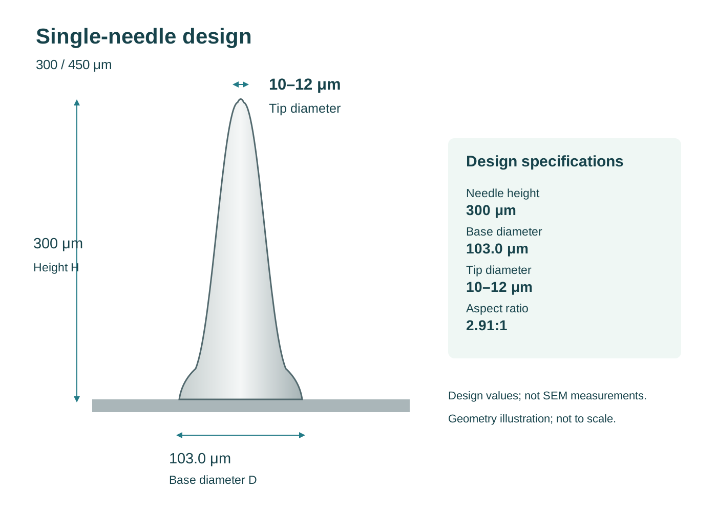
Drawing annotations: H 300 μm, base diameter 103.0 μm, tip diameter 10–12 μm, H/D 2.91:1.
Open original image ↗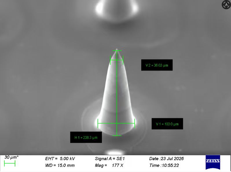
Original annotations: H1 238.3 μm, V1 103.0 μm, V2 36.03 μm.
Open original image ↗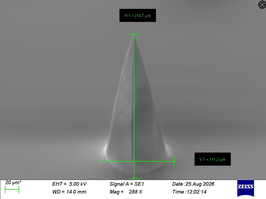
Original annotations: H1 214.7 μm, V1 111.2 μm.
Open original image ↗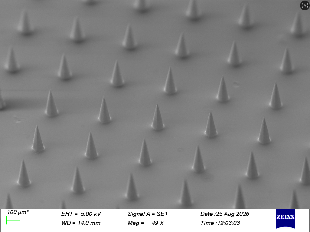
Finished array, with original scale bar and instrument information.
Open original image ↗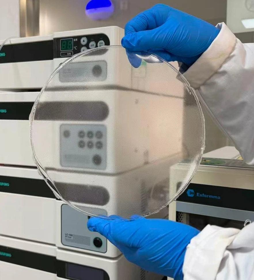
Original product photograph supplied by Novelty.
Open original image ↗Design values and SEM measurements are shown separately. Measurement orientation, sample condition, and tolerances have not yet been documented. No shrinkage rate or acceptance range is inferred.
Needle length 300 μm · Spacing 600 μm
Design length: 300 μm. Original SEM annotations are retained. SEM measurements describe the pictured sample; they do not establish nominal length or skin penetration depth.
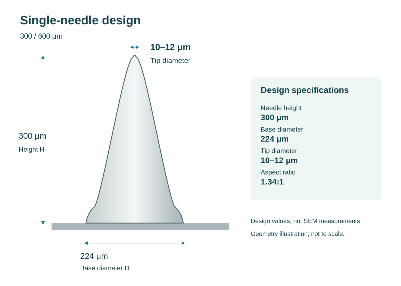
Drawing annotations: H 300 μm, base diameter 224 μm, tip diameter 10–12 μm, H/D 1.34:1; curvature parameters are estimates.
Open original image ↗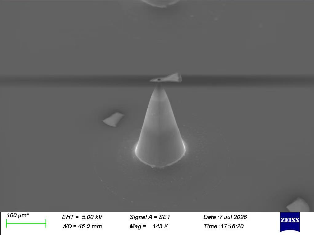
Silicon master needle morphology; original 100 μm scale bar and instrument information retained.
Open original image ↗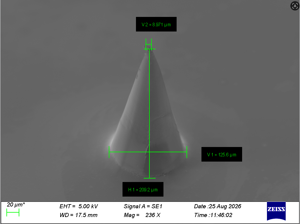
Original annotations: H1 209.2 μm, V1 125.6 μm, V2 8.971 μm.
Open original image ↗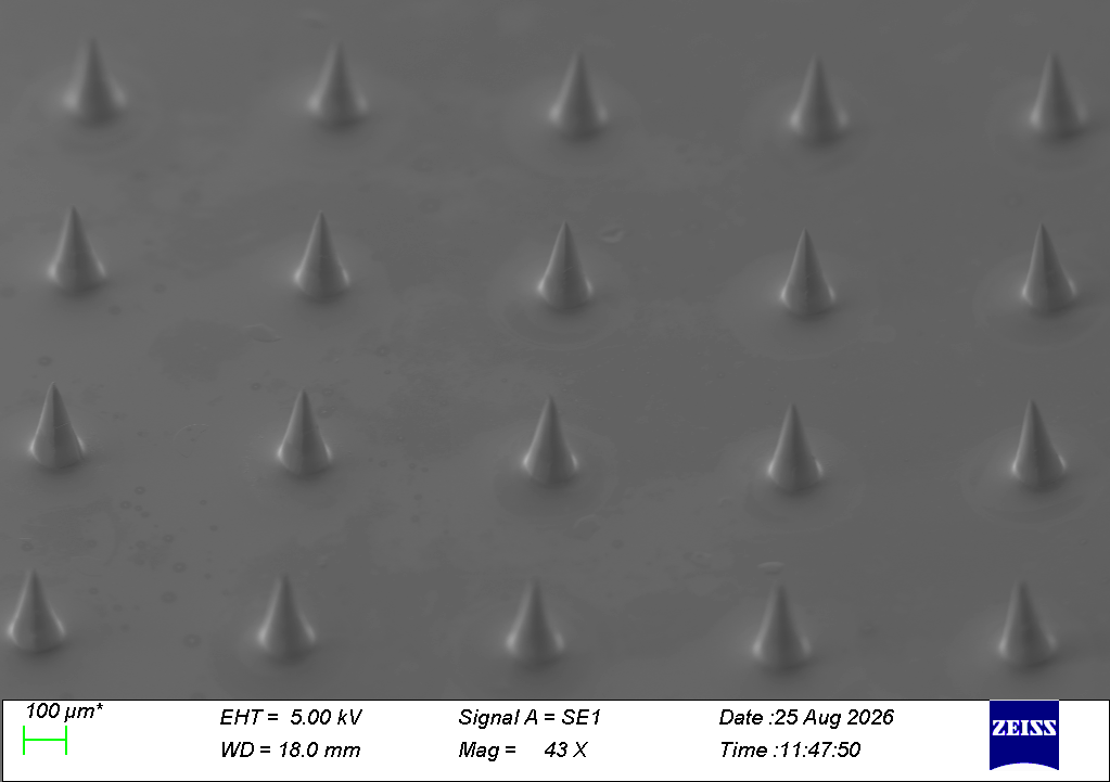
Finished array, with original scale bar and instrument information.
Open original image ↗Design values and SEM measurements are shown separately. Measurement orientation, sample condition, and tolerances have not yet been documented. No shrinkage rate or acceptance range is inferred.
Needle length 600 μm · Spacing 800 μm
Design parameters are updated; microscopy images retain their original measurements. The design needle length is 600 μm; H1 on the SEM image is an image-specific measurement, not nominal needle length or skin penetration depth.
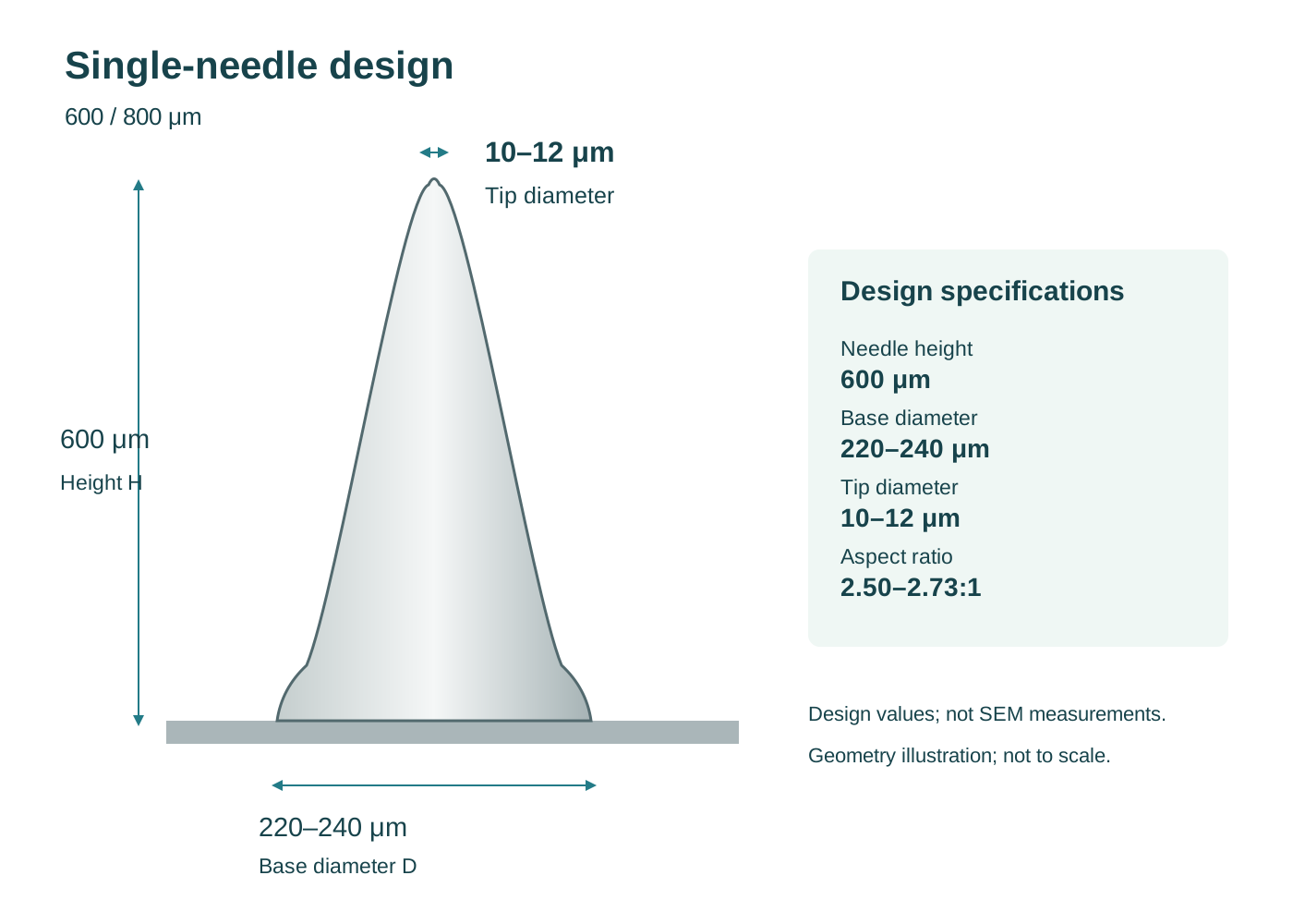
Drawing annotations: H 600 μm, base diameter 220–240 μm, tip diameter 10–12 μm, H/D 2.50–2.73:1; curvature parameters are estimates.
Open original image ↗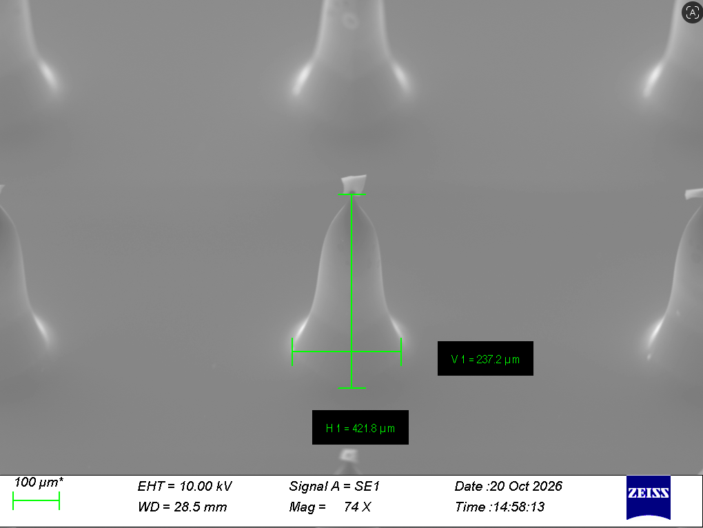
Original annotations: H1 421.8 μm, V1 237.2 μm; scale bar and instrument information retained.
Open original image ↗Design dimensions and SEM measurements are retained separately; measurement orientation, sample condition and specification tolerances remain to be documented.
Needle length 800 μm · Spacing 1000 μm
Design parameters are updated; microscopy images retain their original measurements. The design needle length is 800 μm; H1 on the SEM image is an image-specific measurement, not nominal needle length or skin penetration depth.
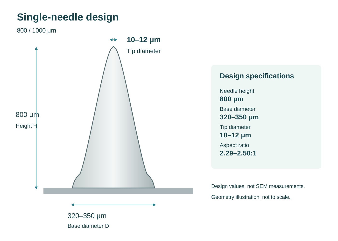
Drawing annotations: H 800 μm, base diameter 320–350 μm, tip diameter 10–12 μm, H/D 2.29–2.50:1; curvature parameters are estimates.
Open original image ↗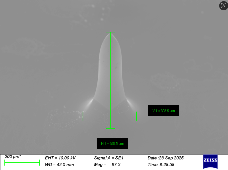
Original annotations: H1 555.5 μm, V1 308.6 μm; scale bar and instrument information retained.
Open original image ↗Design dimensions and SEM measurements are retained separately; measurement orientation, sample condition and specification tolerances remain to be documented.
Follow each stage: needle design, array layout, master fabrication, PDMS molding, microneedle fabrication, and SEM inspection.
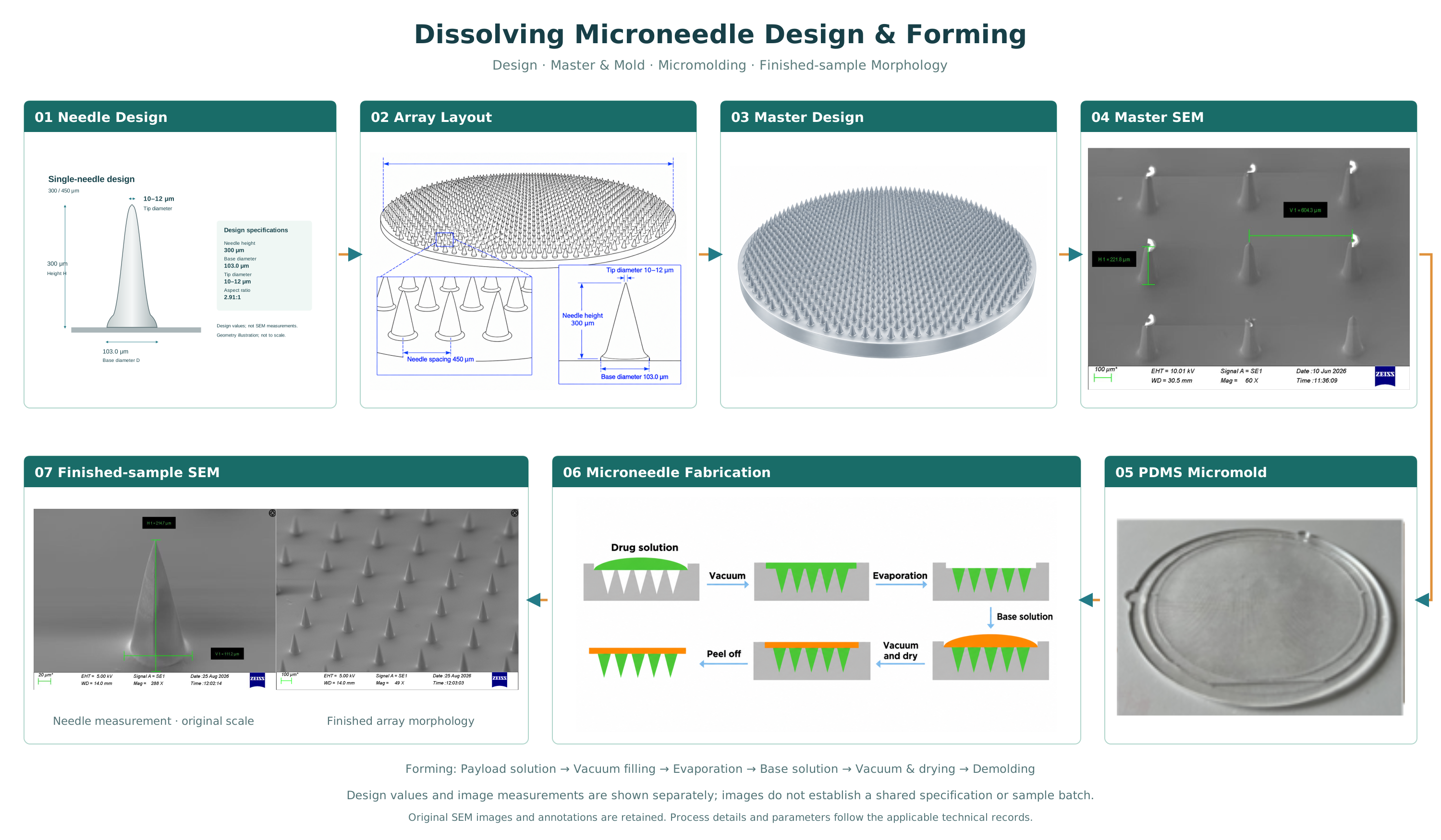
Examine needle shape, dimensions, integrity, and mechanical response under defined conditions.
Measure hydration and dissolution using a specified medium, temperature, and time period.
Assess insertion, ingredient distribution, local tolerance, and usability with methods suited to the product.
Track changes in appearance, structure, and material condition during storage.
Results depend on the sample and test conditions. We identify the method, conditions, and measured outcome so the data can be interpreted in context.
See how Raman signals vary across skin layers from 30 minutes to 6 hours after application, comparing hydrolyzed sodium hyaluronate with its microneedle formulation.
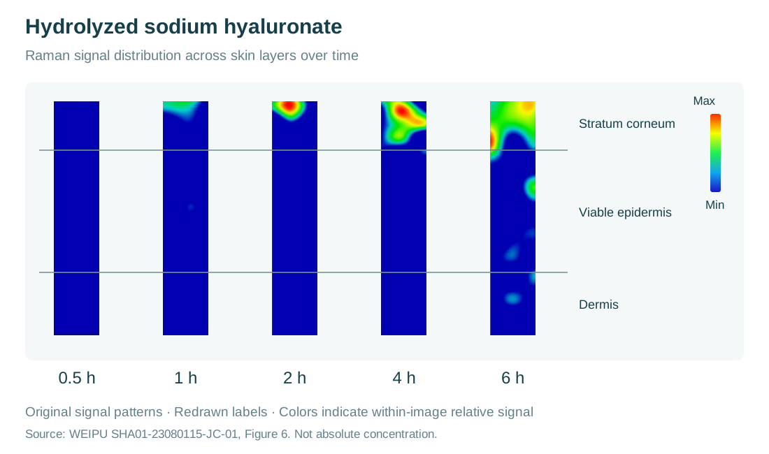
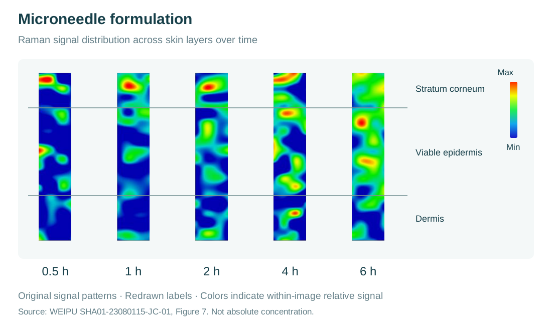
The report describes the non-microneedle sample as concentrated mainly in the stratum corneum at 1, 2, and 4 hours, reaching the viable epidermis by 6 hours. With microneedles, signals were observed in the dermal region at 30 minutes and across all three layers at later time points.
Colors retain the original heatmap scale and show relative Raman signals within each image. They help locate signals across skin layers. The images do not provide a shared absolute concentration calibration or measure needle insertion depth. The topical sample had a reported dermal value of 0.03% at 6 hours.
| Sample | 0.5 h | 1 h | 2 h | 4 h | 6 h |
|---|---|---|---|---|---|
| Hydrolyzed sodium hyaluronate | 0% | 0.15% | 2.08% | 4.74% | 6.12% |
| Microneedle formulation | 6.32% | 9.53% | 11.48% | 16.91% | 22.74% |
| Sample / layer | 0.5 h | 1 h | 2 h | 4 h | 6 h |
|---|---|---|---|---|---|
| Topical · epidermis | 0% | 0.15% | 2.08% | 4.74% | 6.09% |
| Topical · dermis | 0% | 0% | 0% | 0% | 0.03% |
| Microneedles · epidermis | 4.97% | 8.46% | 8.76% | 13.53% | 19.81% |
| Microneedles · dermis | 1.35% | 1.07% | 2.72% | 3.38% | 2.93% |
Study conditions: one participant; forearm; three 1 × 1 cm² areas, with five measurement points per area. Testing used a HORIBA LabRAM Odyssey at 22 ± 2°C and 50 ± 10% relative humidity. Results are specific to this participant, these samples, and these conditions. Relative penetration is not absolute concentration, systemic absorption, or clinical efficacy.
Source: WEIPU report SHA01-23080115-JC-01, September 13, 2023. Methods: pages 4–5; figures: pages 9–10; data: pages 10–11. These excerpts do not replace the complete report.
Formats for the eye area, forehead, between the brows, smile lines, lips, and localized care.
Dissolving microcrystal rollers with handles, designed for a distinct application routine.
A separate product category made to company standards in China. Sleep patch information is available; glucosamine patch details will follow.
Medical devices, pharmaceutical delivery, and animal health each have their own development and testing requirements.
Not necessarily. The application site, material, and method matter. Needle length does not equal insertion depth.
It contains a preformed array of microscopic structures. Product benefits must be established through testing.
Usually only the soluble needles or matrix dissolve. Follow the product instructions for removing the backing and other components.
No. Dissolving needles dissolve; hydrogel-forming needles swell. A companion hydrogel patch is a separate component.
It shows surface structure and dimensions. It does not establish delivery performance or clinical efficacy.
Tell us where and how the product will be used. We can then discuss materials, design, manufacturing, and testing.